Clinical Anatomy Hand Wrist Palmar Aspect Flexors

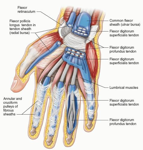
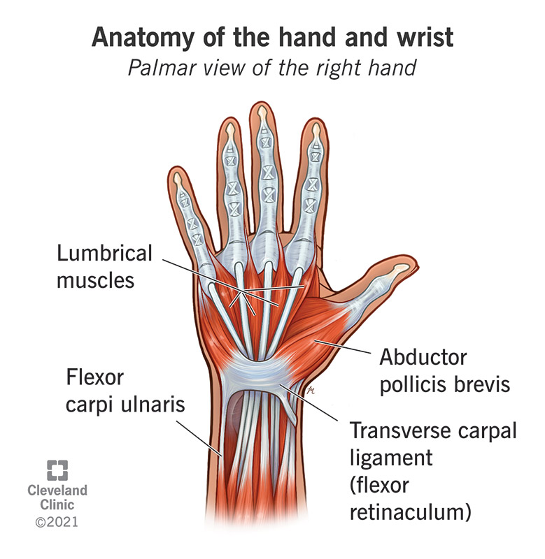
Clinical Anatomy Hand Wrist Palmar Aspect Flexors Youtube Where do i get my information from: armandoh.org resourcehit the like button!facebook: facebook armandohasudungansupport me: www. They control the area of your hand that’s opposite your thumb. interossei muscles: interossei muscles are between the metacarpal bones in your palm. they help your fingers move side to side. lumbrical muscles: lumbrical muscles are at the base of your four non thumb fingers. they help you flex your fingers.

Hand And Wrist Radiology Key Comprehensive anatomy of wrist and hand and common sites of injury. palmar and extensor neurovasculature. the origin, course, distribution, and anastomosis of the branches of the major vessels that supply drain the hand (superficial and deep palmar arches) and fingers (digital branches). the origin, course, and function of the terminal branches. Flexion ( or palmar flexion) at the radioulnar joint is described as the movement in which the palmar aspect of the hand moves towards the forearm in the sagittal plane. during flexion, the scaphoid and lunate bones glide over the concave articular surface of the distal radius in a posterosuperior direction. The hand and wrist have a total of 27 bones arranged to roll, spin and slide [5]; allowing the hand to explore and control the environment and objects. the carpus is formed from eight small bones collectively referred to as the carpal bones. the carpal bones are bound in two groups of four bones: the pisiform, triquetrum, lunate and scaphoid on. The wrist joint (also known as the radiocarpal joint) is an articulation between the radius and the carpal bones of the hand. it is condyloid type synovial joint which marks the area of transition between the forearm and the hand. in this article, we shall look at the anatomy of the wrist joint – its structure, neurovasculature and clinical.

Anatomy Of The Hand Wrist Bones Muscles Ligaments The hand and wrist have a total of 27 bones arranged to roll, spin and slide [5]; allowing the hand to explore and control the environment and objects. the carpus is formed from eight small bones collectively referred to as the carpal bones. the carpal bones are bound in two groups of four bones: the pisiform, triquetrum, lunate and scaphoid on. The wrist joint (also known as the radiocarpal joint) is an articulation between the radius and the carpal bones of the hand. it is condyloid type synovial joint which marks the area of transition between the forearm and the hand. in this article, we shall look at the anatomy of the wrist joint – its structure, neurovasculature and clinical. Introduction. the wrist includes three joints: the distal radioulnar joint, the radiocarpal joint and the midcarpal joint. the movements at the wrist are flexion and extension, radial and ulnar deviation and pronation and supination (at the distal radioulnar joint). optimal wrist function requires adequate range of motion. The scapholunate (sl) ligament is located about 1 cm distal to lister’s tubercle. figure 61.1 a and b, surface anatomy of the hand and wrist. the dotted line represents the original description of kaplan’s cardinal line. the dashed line denotes the distal palmar crease. on the palmar view, the superficial flexor carpi radialis (fcr) and.

Comments are closed.