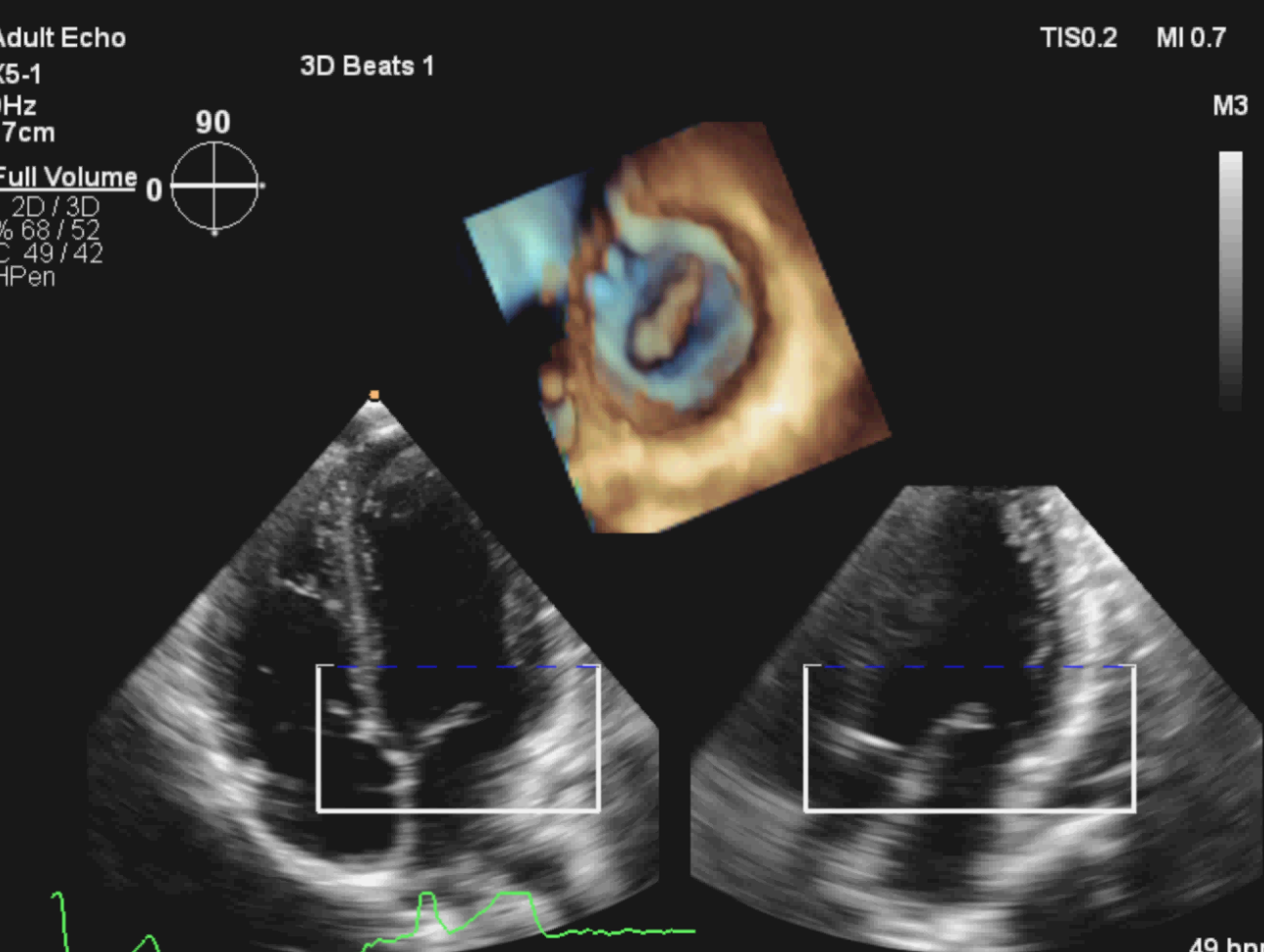
3d Echocardiogram Diameter And Ventricular Function Are Normal With

3d Echocardiogram Diameter And Ventricular Function Are Normal With Introduction. impressive technological advancements have made 3d echocardiography (3de) a fundamental tool for the assessment of left ventricular (lv) volumes and function. 1 as 3de does not rely on geometric assumptions about lv shape and allows assessment of lv volumes, lv ejection fraction, and all strain components within a single data set acquisition, it has overcome most of the 2d. With regards 3d volumetric assessment and 3d derived ejection fraction, the bse stresses that reference intervals for 2d derived ejection fraction do not apply to 3d results: for example, an ejection fraction of 56% derived using 3d software is not necessarily normal, and comparison with vendor specific reference intervals should be used.

3d Echocardiography вђ Thinking 3d Normal 2d measurements from the apical 4 chamber view; rv medio lateral end diastolic dimension ≤ 4.3 cm, rv end diastolic area ≤ 35.5 cm 2 (89). § at a nyquist limit of 50 60 cm s. φ cut off values for regurgitant volume and fraction are not well validated. † steep deceleration is not specific for severe pr. Echocardiography remains the most widely used modality to assess left ventricular (lv) chamber size and function. currently this assessment is most frequently performed using two dimensional (2d) echocardiography. however, three dimensional (3d) echocardiography has been shown to be more accurate and reproducible than 2d echocardiography. current normative reference values for 3d lv analysis. The world alliance societies of echocardiography study was designed to sample normal subjects from around the world to provide more universal global reference ranges. the aim of this study was to assess the worldwide feasibility of lv 3d echocardiography and report on size and functional measurements. Aim: to obtain the normal ranges for 3d echocardiography (3de) measurement of left ventricular (lv) volumes, function, and strain from a large group of healthy volunteers.

Comments are closed.