Benign Liver Mass Ohsu Contrast Enhanced Ultrasound Ceus

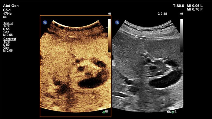
Benign Liver Mass Ohsu Contrast Enhanced Ultrasound Ceus Youtube Ceus movie of the liver. split screen image with contrast image on the right and standard ultrasound on the left. transverse view of the right lobe of the. A total of 1328 focal liver lesions (755 malignant and 573 benign) were assessed. the reference standard diagnosis was made by means of liver biopsy in 75% of cases and by contrast enhanced ct or contrast enhanced mri in the other cases. the accuracy of ceus for the diagnosis of focal liver lesions was 90.3%.

Contrast Enhanced Ultrasound Of Focal Liver Masses Contrast agent–enhanced ultrasonography of liver lesions is both accurate and reproducible for evaluation of benign and malignant liver tumors. use of an imaging algorithm and a controlled sonographic technique, including dedicated arterial phase cine imaging and imaging every 30 seconds in the portal venous phase and the delayed (or late. It is estimated that the cost of the ceus examination, if performed during the same sitting as a b mode ultrasound, is £65; a contrast ct examination would cost between £116 and £162 and a contrast mr examination between £189 and £366. 3. nice guidelines state that 75% of incidentally detected liver lesions are benign. Our previously published educational article, “contrast enhanced us approach to the diagnosis of focal liver masses,” describes a practical algorithmic approach to imaging liver lesions with contrast agent–enhanced us (ceus) . it focuses on the evaluation of all nodules in all adult patients. Ceus of the liver becomes even more relevant with the recent fda approval of a second generation ultrasound contrast agent lumason (bracco diagnostics inc., monroe township, nj) for liver imaging, making focal liver lesion characterization with ceus the obvious next step after lesion detection on surveillance ultrasound.

Ceus Contrast Enhanced Ultrasound Features Aixplorer Mach Home Our previously published educational article, “contrast enhanced us approach to the diagnosis of focal liver masses,” describes a practical algorithmic approach to imaging liver lesions with contrast agent–enhanced us (ceus) . it focuses on the evaluation of all nodules in all adult patients. Ceus of the liver becomes even more relevant with the recent fda approval of a second generation ultrasound contrast agent lumason (bracco diagnostics inc., monroe township, nj) for liver imaging, making focal liver lesion characterization with ceus the obvious next step after lesion detection on surveillance ultrasound. Contrast enhanced ultrasound (ceus) liver imaging reporting and data system (li rads) 2017 – a review of important differences compared to the ct mri system tae kyoung kim , 1 seung yeon noh , 2 stephanie r wilson , 3 yuko kono , 4 fabio piscaglia , 5 hyun jung jang , 1 andrej lyshchik , 6 christoph f. dietrich , 7 juergen k. willmann , 8. Contrast enhanced ultrasound (ceus) continues to gain traction as a technique that complements traditional b mode and doppler ultrasound in the evaluation of the liver and other organs. because the micro vasculature can be visualized with ceus and real time imaging of tissue perfusion can be performed, imaging with this technique yields.

Ceus Contrast Enhanced Ultrasound Philips Healthcare Contrast enhanced ultrasound (ceus) liver imaging reporting and data system (li rads) 2017 – a review of important differences compared to the ct mri system tae kyoung kim , 1 seung yeon noh , 2 stephanie r wilson , 3 yuko kono , 4 fabio piscaglia , 5 hyun jung jang , 1 andrej lyshchik , 6 christoph f. dietrich , 7 juergen k. willmann , 8. Contrast enhanced ultrasound (ceus) continues to gain traction as a technique that complements traditional b mode and doppler ultrasound in the evaluation of the liver and other organs. because the micro vasculature can be visualized with ceus and real time imaging of tissue perfusion can be performed, imaging with this technique yields.

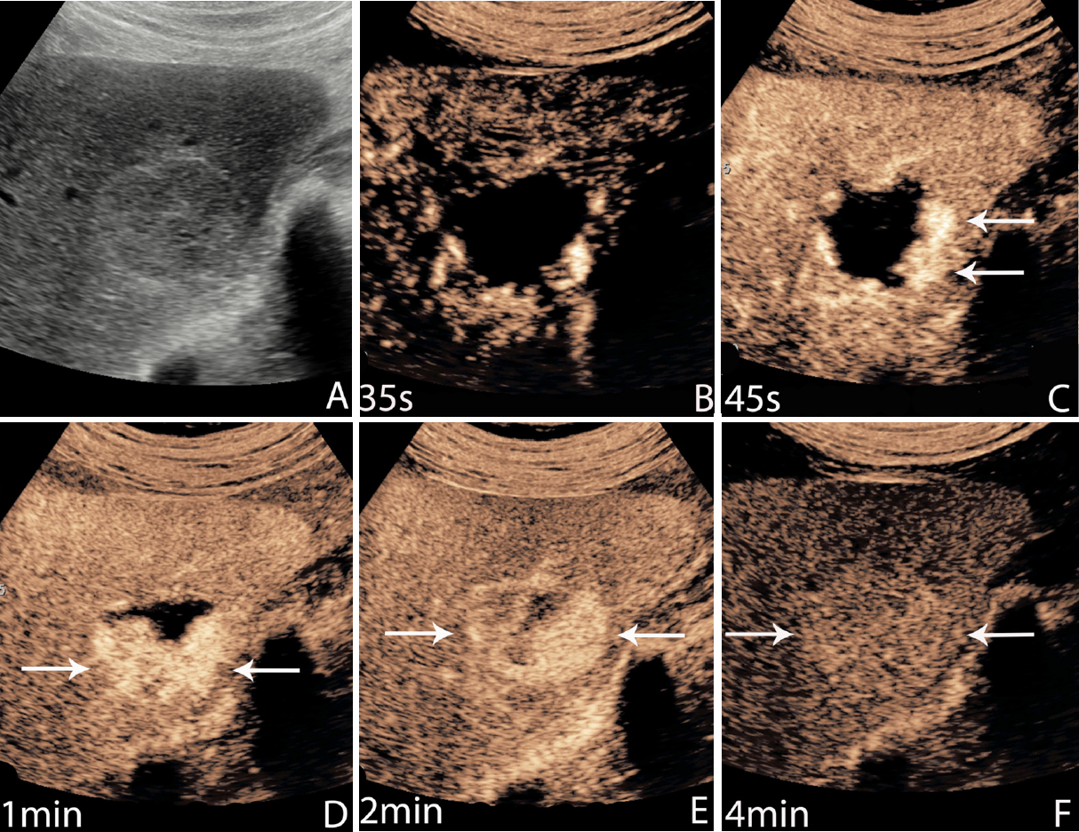
Contrast Enhanced Ultrasound Ceus Showing Intense Peripheral

Comments are closed.