Colles Fracture вђ Medicolearning

Colles Fractures Central Coast Orthopedic Medical Group Colles fracture. post category: flashcard orthopedics; post published: february 2, 2022; colles fracture. share share this content. opens in a new window;. Named after abraham colles, who first described a distal radius fracture in 1814 at the royal college of surgeons in dublin, the colles fracture is one of the most common fractures encountered in orthopedic practice representing 17.5 % (one sixth) of all adult fractures presenting to the emergency department.[1][2] the colles fracture is defined as a distal radius fracture with dorsal.

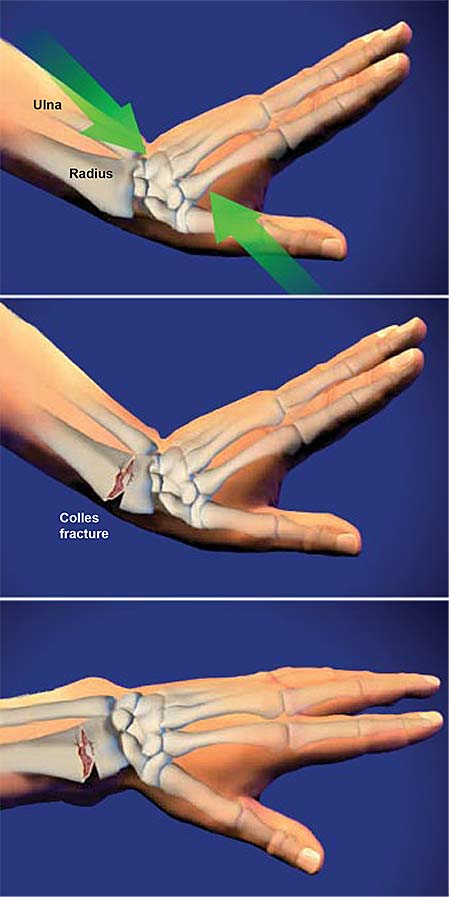
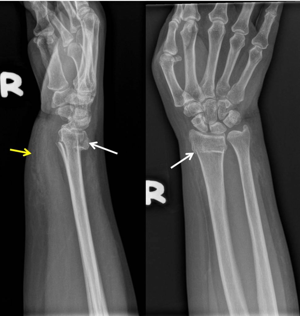
Colles Fracture вђ Medicolearning Colles' fractures are a common presentation to emergency departments across the globe. the eponymous fracture is a dorsally angulated extra articular distal radial metaphyseal single segment fracture. everything else is a distal radial fracture or a smiths or a bartons or a chauffeur fracture or galeazzi. Colles fractures are very common extra articular fractures of the distal radius that occur as the result of a fall onto an outstretched hand. they consist of a fracture of the distal radial metaphyseal region with dorsal angulation and impaction, but without the involvement of the articular surface. this article describes radiographic features. The colles fracture, the most common fracture of the wrist, was first described by abraham colles in 1814. in this injury, there is a complete fracture of the distal radius (typically the last two centimeters) usually accompanied by damage to the ulnar collateral ligament or the ulnar styloid process. middle aged women and elderly are more. Carpal tunnel syndrome, which is a compression of the nerves in your wrist. symptoms include pain, numbness and weakness in your wrist and hand. reflex sympathetic dystrophy causes burning pain in your arms, legs, hands or feet. secondary osteoarthritis might develop in your wrist. symptoms include pain and swelling of a deformed joint.

Colles Fracture вђ Radiology Cases The colles fracture, the most common fracture of the wrist, was first described by abraham colles in 1814. in this injury, there is a complete fracture of the distal radius (typically the last two centimeters) usually accompanied by damage to the ulnar collateral ligament or the ulnar styloid process. middle aged women and elderly are more. Carpal tunnel syndrome, which is a compression of the nerves in your wrist. symptoms include pain, numbness and weakness in your wrist and hand. reflex sympathetic dystrophy causes burning pain in your arms, legs, hands or feet. secondary osteoarthritis might develop in your wrist. symptoms include pain and swelling of a deformed joint. A colles fracture is a complete fracture of the radius bone of the forearm close to the wrist resulting in an upward (posterior) displacement of the radius and obvious deformity. it is commonly called a “broken wrist” in spite of the fact that the distal radius is the location of the fracture, not the carpal bones of the wrist. [1]. 5mm shortening of the radius. stable fracture. use procedural sedation or hematoma block. hang 10 lb weight with finger traps or otherwise provide longitudinal traction. recreate the injury by extending wrist to 90 degrees while elbow is flexed. pull distal segment back, up, and then out; use both thumbs to apply volar pressure.

Comments are closed.