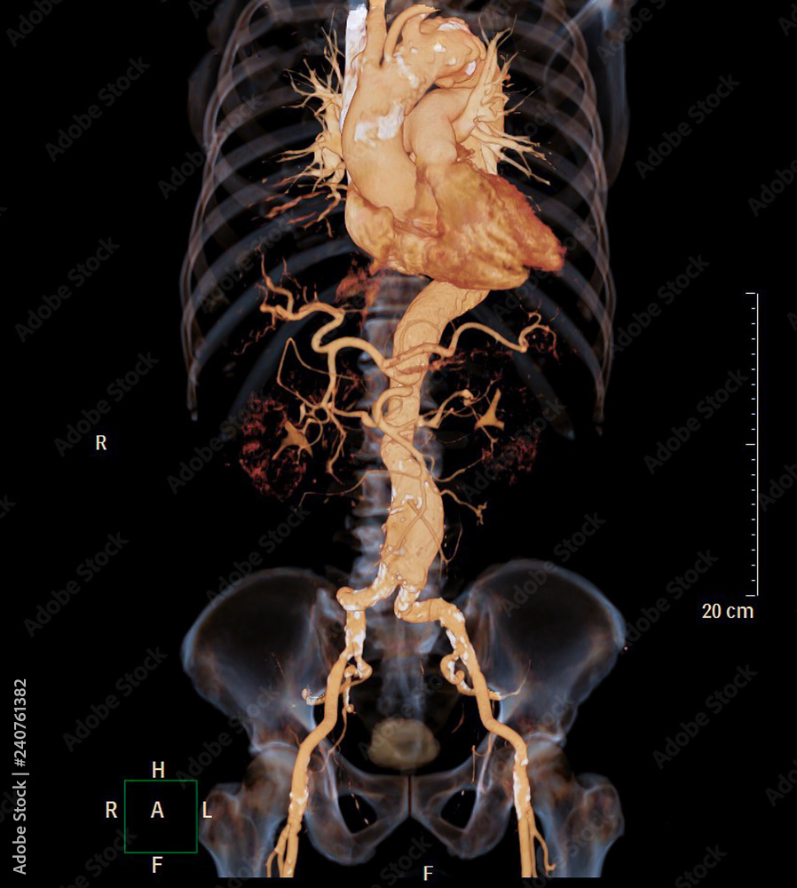
Ct Angiography Of Abdominal Aorta 3d Rendering Image With X Ray Image

Ct Angiography Of Abdominal Aorta 3d Rendering Image With X Ray Image The indications for a ct of the abdominal aorta vary depending on an emergency versus outpatient presentation 1. generally, the abdominal aorta is included in standard trauma imaging (chest abdomen pelvis), which includes an arterial chest and portal venous abdomen. thus, specific abdominal aortic imaging is only requested when high suspicion. Objective. the purpose of this article is to present an overview of cinematic rendering, illustrating its potential advantages and applications. conclusion. volume rendered reconstruction, obtaining 3d visualization from original ct datasets, is increasingly used by physicians and medical educators in various clinical and educational scenarios. cinematic rendering is a novel 3d rendering.

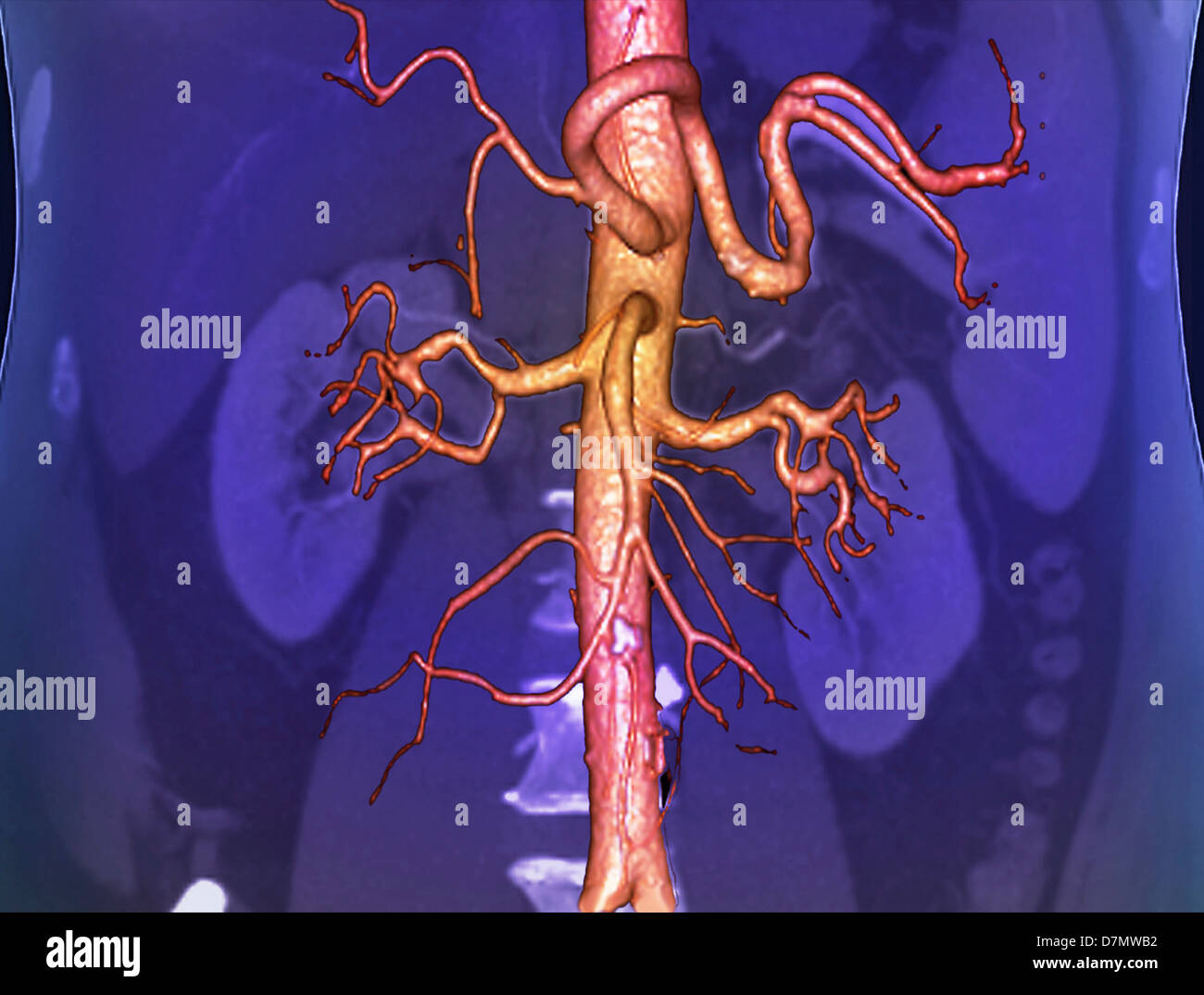
Ct Angiography Of Abdominal Aorta 3d Rendering Image With X Ray Image Purpose aorta segmentation is extremely useful in clinical practice, allowing the diagnosis of numerous pathologies, such as dissections, aneurysms and occlusive disease. in such cases, image segmentation is prerequisite for applying diagnostic algorithms, which in turn allow the prediction of possible complications and enable risk assessment, which is crucial in saving lives. the aim of this. Ct angiography provides excellent anatomical details of abdominal aorta and stent grafts, thus enabling assessment of the diameter of aneurysms and stent grafts relative to the aortic branches. despite these advantages, ct angiography is limited to image visualization and does not provide information about hemodynamic changes to the abdominal. Computed tomographic (ct) angiography (cta) has become the preferred imaging test of choice for various aortic conditions because of its excellent spatial resolution, rapid image acquisition, and its wide availability. cta provides a robust tool for planning aortic interventions and diagnosing acute and chronic vascular diseases in the abdomen. The 3d volume rendering image shows a complete obstruction of the subrenal aorta and iliac bifurcation. the vascularization of the internal and external iliac arteries and the common femoral.

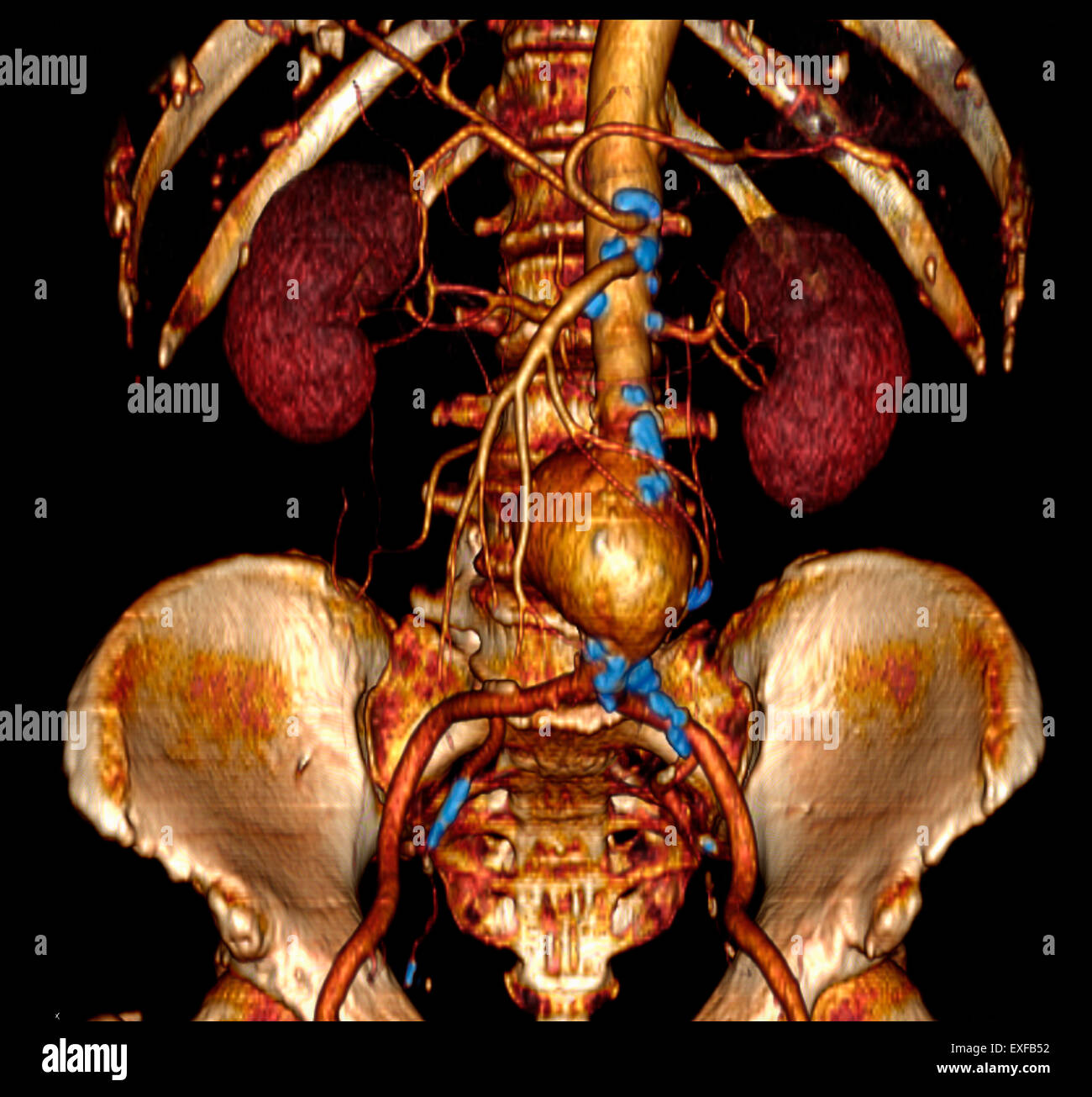
3d Abdominal Angiographic Ct Scan Abdominal Aortic Aneurysm Stoc Computed tomographic (ct) angiography (cta) has become the preferred imaging test of choice for various aortic conditions because of its excellent spatial resolution, rapid image acquisition, and its wide availability. cta provides a robust tool for planning aortic interventions and diagnosing acute and chronic vascular diseases in the abdomen. The 3d volume rendering image shows a complete obstruction of the subrenal aorta and iliac bifurcation. the vascularization of the internal and external iliac arteries and the common femoral. The introduction and widespread availability of 16 section multi–detector row computed tomographic (ct) technology and, more recently, 64 section scanners, has greatly advanced the role of ct angiography in clinical practice. ct angiography has become a key component of state of the art imaging, with applications ranging from oncology (eg, staging of pancreatic or renal cancer) to classic. Objective. the two most widely used postprocessing 3d tools in clinical practice are volume rendering (vr) and maximum intensity projection (mip). with the use of current generation mdct, these techniques enable accurate characterization of arterial anatomy and pathology in all anatomic regions. recently, the vr algorithm has been enhanced by the incorporation of a new lighting model. this new.

Abdominal Aorta 3d Ct Scan Stock Photo 56392646 Alamy The introduction and widespread availability of 16 section multi–detector row computed tomographic (ct) technology and, more recently, 64 section scanners, has greatly advanced the role of ct angiography in clinical practice. ct angiography has become a key component of state of the art imaging, with applications ranging from oncology (eg, staging of pancreatic or renal cancer) to classic. Objective. the two most widely used postprocessing 3d tools in clinical practice are volume rendering (vr) and maximum intensity projection (mip). with the use of current generation mdct, these techniques enable accurate characterization of arterial anatomy and pathology in all anatomic regions. recently, the vr algorithm has been enhanced by the incorporation of a new lighting model. this new.

Comments are closed.