Didelphys Uterus Ultrasound

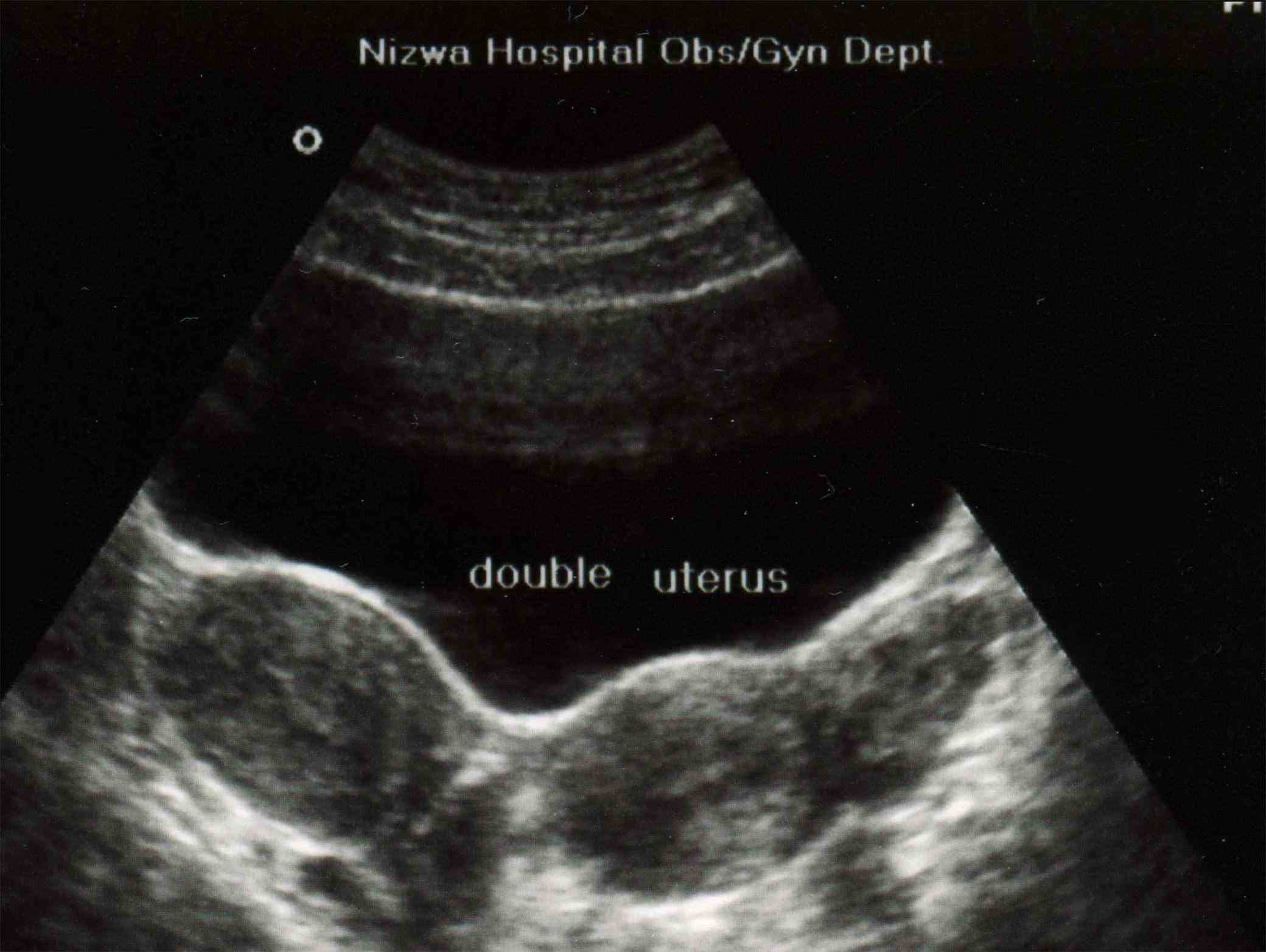
Didelphys Uterus Ultrasound Uterus didelphys is a müllerian duct anomaly where there are two separate uterine horns and cervices. ultrasound shows two divergent uterine bodies with a large fundal cleft and no communication between them. A uterus didelphys is a type of müllerian duct anomaly (class iii) where there is complete duplication of uterine horns as well as duplication of the cervix, with no communication between them. it results from failed ductal fusion that occurs between the 12 th and 16 th week of pregnancy and is characterized by two symmetric, widely divergent uterine horns and two cervixes.

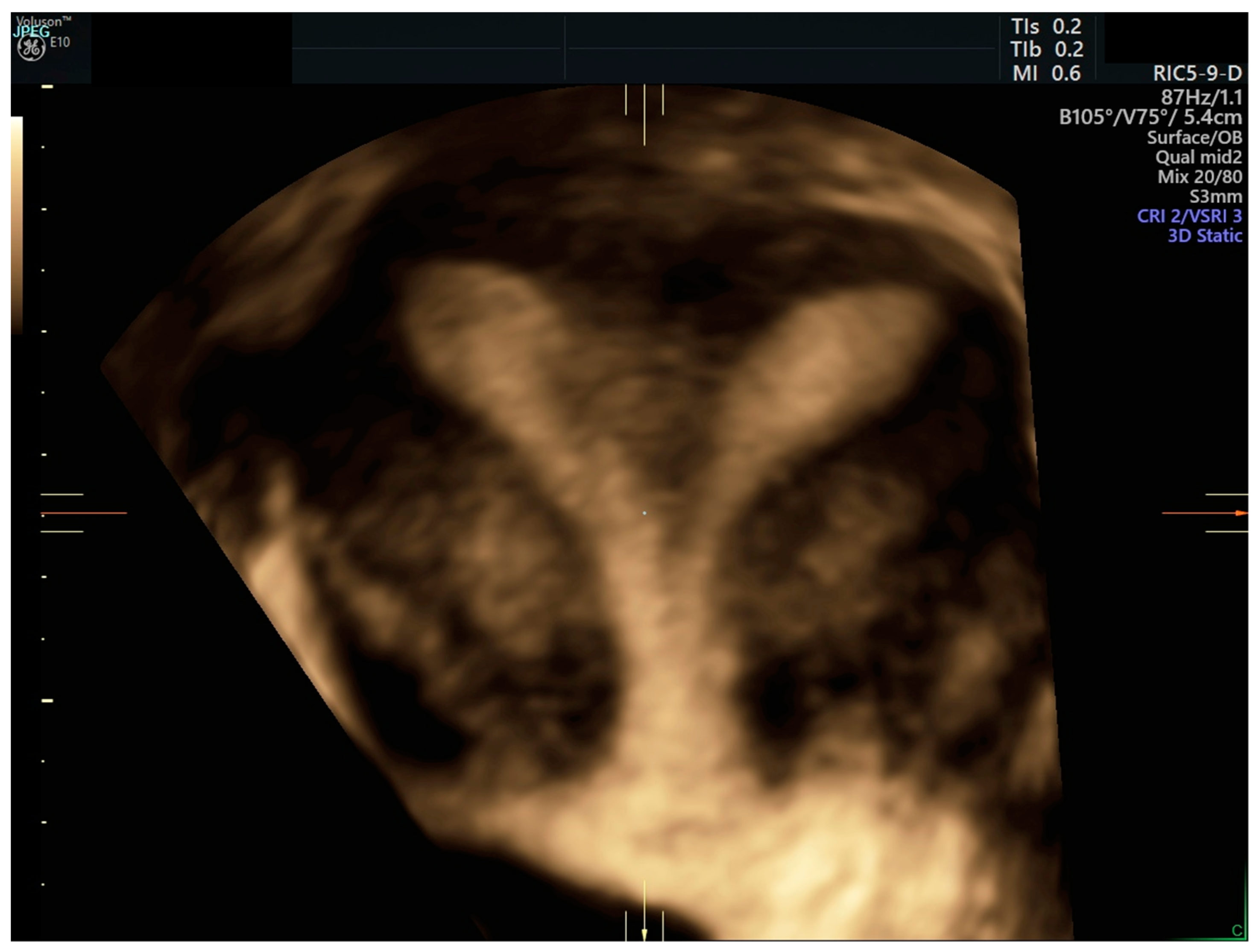
Didelphys Uterus Ultrasound Uterus didelphys is a rare condition where you’re born with two uteruses. learn how it affects your periods, pregnancy and sex life, and how it’s diagnosed with ultrasound and other tests. A healthy 28 year old primigravida, who presented for pregnancy dating ultrasound at 12 weeks gestation, was incidentally diagnosed with a didelphys uterus. transabdominal ultrasound identified a viable intrauterine pregnancy in the right horn and both uteri were confluent with a single cervix (figure 2). both kidneys were present and appeared. Uterus didelphys showing in 3d ultrasound mode. confirmation of the diagnosis was made possible by hysteroscopy, during which a double uterus, two cervixes, and an elongated septum running through half of the vagina were visualized. Specialty. gynecology. uterus didelphys (from ancient greek di 'two' and delphus 'womb'; sometimes also uterus didelphis) represents a uterine malformation where the uterus is present as a paired organ when the embryogenetic fusion of the müllerian ducts fails to occur. as a result, there is a double uterus with two separate cervices, and.
Didelphys Uterus Ultrasound Uterus didelphys showing in 3d ultrasound mode. confirmation of the diagnosis was made possible by hysteroscopy, during which a double uterus, two cervixes, and an elongated septum running through half of the vagina were visualized. Specialty. gynecology. uterus didelphys (from ancient greek di 'two' and delphus 'womb'; sometimes also uterus didelphis) represents a uterine malformation where the uterus is present as a paired organ when the embryogenetic fusion of the müllerian ducts fails to occur. as a result, there is a double uterus with two separate cervices, and. Uterus didelphys is a class iii müllerian anomaly based on the asrm classification. this disorder accounts for approximately 5% of mdas. uterus didelphys occurs as a result of a complete or near complete lack of müllerian duct fusion. each duct develops into an independent hemiuterus and cervix, although partial cervical fusion is generally seen. Initial screening with 2d ultrasound may differentiate septate from didelphys, as the uterine horns are usually disparate with didelphic uterus. if there is a single cervix on exam, then uterus didelphys is unlikely unless the vaginal septum and second vaginal canal was missed.

Figure Transabdominal Ultrasound Of Uterine Didelphys Uterus didelphys is a class iii müllerian anomaly based on the asrm classification. this disorder accounts for approximately 5% of mdas. uterus didelphys occurs as a result of a complete or near complete lack of müllerian duct fusion. each duct develops into an independent hemiuterus and cervix, although partial cervical fusion is generally seen. Initial screening with 2d ultrasound may differentiate septate from didelphys, as the uterine horns are usually disparate with didelphic uterus. if there is a single cervix on exam, then uterus didelphys is unlikely unless the vaginal septum and second vaginal canal was missed.

Comments are closed.