Eye Diagram Labeled Anatomy And Biology
/GettyImages-695204442-b9320f82932c49bcac765167b95f4af6.jpg)
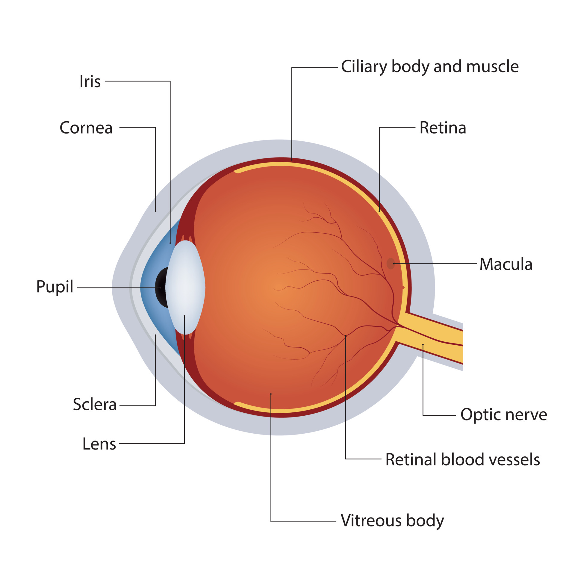
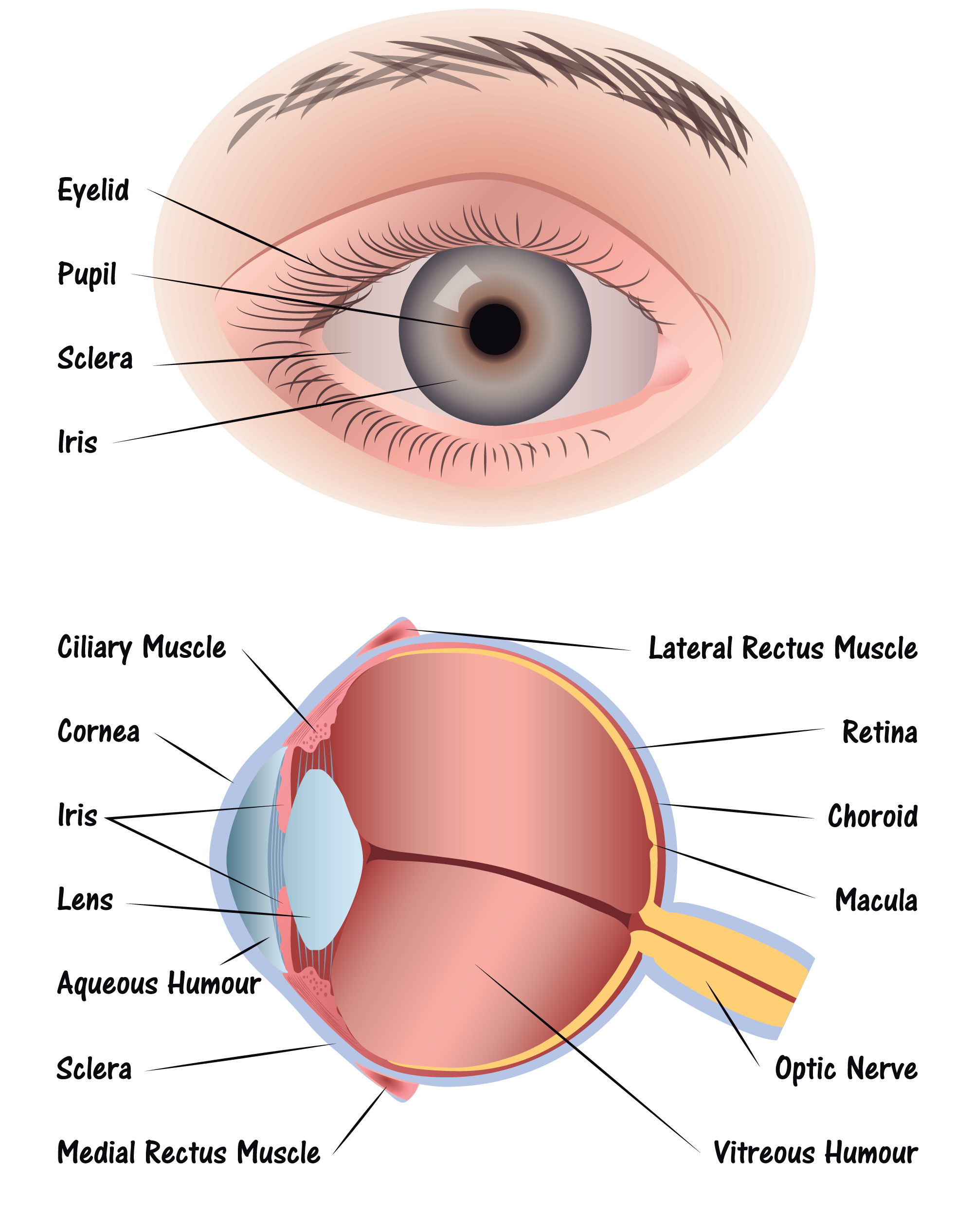
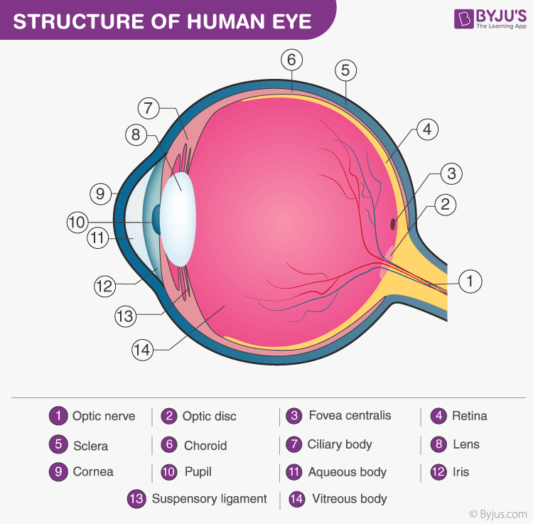
33 Label Of The Eye Labels 2021 The surface of the eye and the inner surface of the eyelids are covered with a clear membrane called the conjunctiva. the layers of the tear film keep the front of the eye lubricated. tears lubricate the eye and are made up of three layers. these three layers together are called the tear film. the mucous layer is made by the conjunctiva. The main function is to refract the light along with the lens. iris: it is the pigmented, coloured portion of the eye, visible externally. the main function of the iris is to control the diameter of the pupil according to the light source. pupil: it is the small aperture located in the centre of the iris.

Structure Of Anatomy Human Eye Detailed Diagram Of Eyeball Side View The following are parts of the human eyes and their functions: 1. conjunctiva. the conjunctiva is the membrane covering the sclera (white portion of your eye). the conjunctiva also covers the interior of your eyelids. conjunctivitis, often known as pink eye, occurs when this thin membrane becomes inflamed or swollen. Labelling the eye. use this interactive to label different parts of the human eye. drag and drop the text labels onto the boxes next to the diagram. selecting or hovering over a box will highlight each area in the diagram. the human eye has several structures that enable entering light energy to be converted to electrochemical energy. Human eye, specialized sense organ in humans that is capable of receiving visual images, which are relayed to the brain. the anatomy of the eye includes auxiliary structures, such as the bony eye socket and extraocular muscles, as well as the structures of the eye itself, such as the lens and the retina. Bony cavity within the skull that houses the eye and its associated structures (muscles of the eye, eyelid, periorbital fat, lacrimal apparatus) bones of the orbit. maxilla, zygomatic bone, frontal bone, ethmoid bone, lacrimal bone, sphenoid bone and palatine bone. structure of the eye. cornea, anterior chamber, lens, vitreous chamber and retina.

Eye Diagram Discovery Eye Foundation Human eye, specialized sense organ in humans that is capable of receiving visual images, which are relayed to the brain. the anatomy of the eye includes auxiliary structures, such as the bony eye socket and extraocular muscles, as well as the structures of the eye itself, such as the lens and the retina. Bony cavity within the skull that houses the eye and its associated structures (muscles of the eye, eyelid, periorbital fat, lacrimal apparatus) bones of the orbit. maxilla, zygomatic bone, frontal bone, ethmoid bone, lacrimal bone, sphenoid bone and palatine bone. structure of the eye. cornea, anterior chamber, lens, vitreous chamber and retina. Near the front of the eye, in the area protected by the eyelids, the sclera is covered by a thin, transparent membrane (conjunctiva), which runs to the edge of the cornea. the conjunctiva also covers the moist back surface of the eyelids and eyeballs. light enters the eye through the cornea, the clear, curved layer in front of the iris and. Diagram of the eye. study the diagram below or click here for an interactive study guide and game! cornea: curved to bend light into your eye, its tough and clear like a windshield to protect your eye from dust. pupil: a hole in the middle of the iris that changes size to let in more or less light. iris: the colored part of the eye with two.

Structure And Functions Of Human Eye With Labelled Diagram Near the front of the eye, in the area protected by the eyelids, the sclera is covered by a thin, transparent membrane (conjunctiva), which runs to the edge of the cornea. the conjunctiva also covers the moist back surface of the eyelids and eyeballs. light enters the eye through the cornea, the clear, curved layer in front of the iris and. Diagram of the eye. study the diagram below or click here for an interactive study guide and game! cornea: curved to bend light into your eye, its tough and clear like a windshield to protect your eye from dust. pupil: a hole in the middle of the iris that changes size to let in more or less light. iris: the colored part of the eye with two.

Comments are closed.