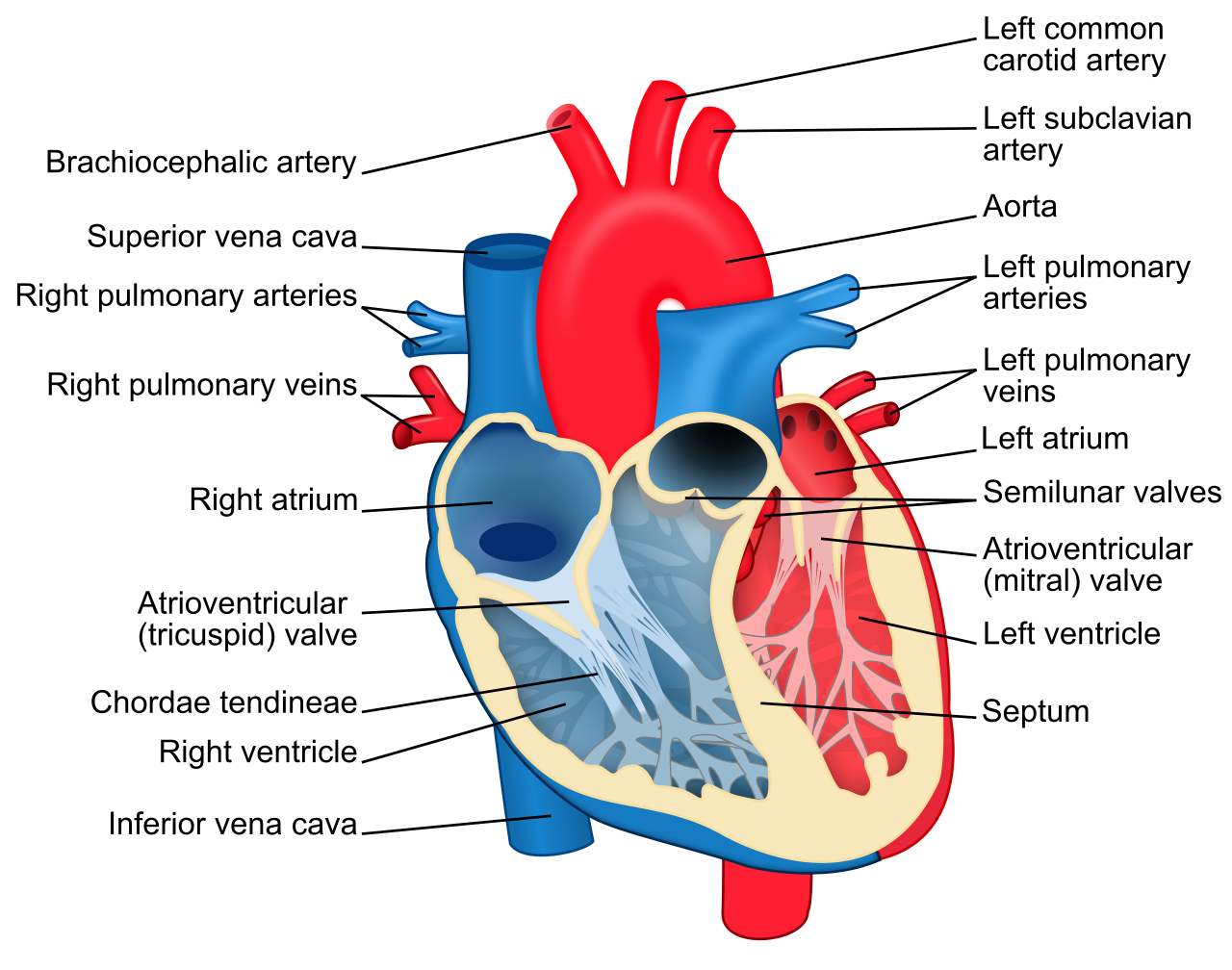
Heart Labeling Diagram

File Heart Diagram En Svg Wikipedia In this interactive, you can label parts of the human heart. drag and drop the text labels onto the boxes next to the heart diagram. if you want to redo an answer, click on the box and the answer will go back to the top so you can move it to another box. Anatomy online heart labeling quiz. the aorta is the main and largest artery in the human body. it originates from the left ventricle. the aortic valve lies at the base of the aorta. it permits blood blood to leave the left ventricle as it contracts. the inferior vena cava and superior vena cava carry blood to the right atrium.

Labeling Diagrams Of The Heart Function and anatomy of the heart made easy using labeled diagrams of cardiac structures and blood flow through the atria, ventricles, valves, aorta, pulmonary arteries veins, superior inferior vena cava, and chambers. includes an exercise, review worksheet, quiz, and model drawing of an anterior view (frontal section) of the heart in order to. The heart is located in the thoracic cavity medial to the lungs and posterior to the sternum. on its superior end, the base of the heart is attached to the aorta, pulmonary arteries and veins, and the vena cava. the inferior tip of the heart, known as the apex, rests just superior to the diaphragm. the base of the heart is located along the. The aortic semilunar valve is between the left ventricle and the opening of the aorta. it has three semilunar cusps leaflets: left left coronary, right right coronary, and posterior non coronary. in clinical practice, the heart valves can be auscultated, usually by using a stethoscope. Heart anatomy in basic terms. the heart is a crucial organ that functions as the body's pump, ensuring blood circulation throughout the body. it consists of four main chambers: left and right atria (upper chambers) left and right ventricles (lower chambers) these chambers work in a coordinated manner to receive oxygen poor blood, pump it to the.

Comments are closed.