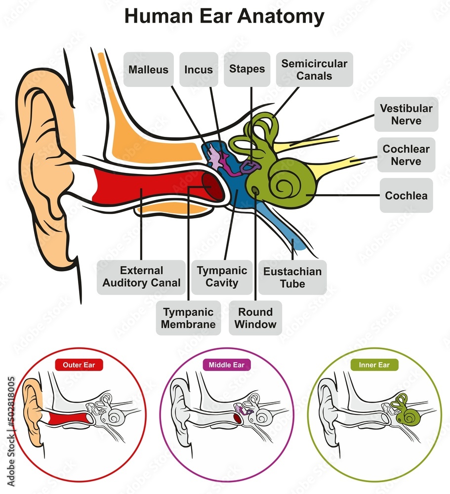
Human Ear Anatomy Infographic Diagram Outer Middle And Innerо

Parts Of The Ear Diagram Labeled The ear is a complex part of an even more complex sensory system. it is situated bilaterally on the human skull, at the same level as the nose. the main functions of the ear are, of course, hearing, as well as constantly maintaining balance. the ear is anatomically divided into three portions: external ear. middle ear. Inner ear: the inner ear, also called the labyrinth, operates the body’s sense of balance and contains the hearing organ. a bony casing houses a complex system of membranous cells. the inner ear.

Outer Ear Anatomy Outer Ear Infection Pain Causes Treatment The ear anatomy consists of three parts: the outer ear, middle ear, and inner ear. the outer ear is the part you can see, including the flap of skin called the pinna and the tube like ear canal. the middle ear is inside your head, and there is a space called the middle ear. it has three tiny bones called ossicles and a cavity called the. Diagnosis. the outer ear is one of three parts of the ear alongside the middle ear (which receives sound waves from the outer ear) and the inner ear (which transmits sound waves to the brain). these sections work together to enable hearing and maintain balance and equilibrium. the outer ear is where sound waves from the air are received. Image size: 35.2 mpixels (101 mb uncompressed) 6250x5625 pixels (20.8x18.8 in 52.9x47.6 cm at 300 ppi). The outer ear is situated superficially next to several bony landmarks. it is posterior to the zygomatic process of the temporal bone as well as the proximal part of the mandibular process and the auricular surface of the mandibular notch. superior to it is the squamous part of the temporal bone, while the styloid process is located inferiorly.

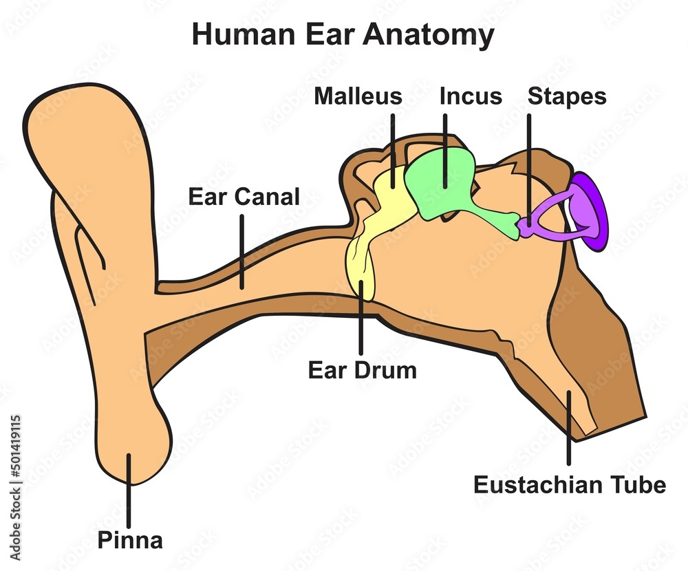
Human Ear Anatomy Infographic Diagram Structure Of Inner Midd Image size: 35.2 mpixels (101 mb uncompressed) 6250x5625 pixels (20.8x18.8 in 52.9x47.6 cm at 300 ppi). The outer ear is situated superficially next to several bony landmarks. it is posterior to the zygomatic process of the temporal bone as well as the proximal part of the mandibular process and the auricular surface of the mandibular notch. superior to it is the squamous part of the temporal bone, while the styloid process is located inferiorly. Your ears have two main functions: hearing and balance. hearing: when sound waves enter your ear canal, your tympanic membrane (eardrum) vibrates. this vibration passes on to three tiny bones (ossicles) in your middle ear. the ossicles amplify and transmit these sound waves to your inner ear. once the sound waves reach your inner ear, tiny hair. The ear canal, which is part of the outer ear, is a tube that connects the cartilage on the outside of the ear to the eardrum. many conditions can affect the ear canal, including infections.

Human Ear Anatomy Infographic Diagram Outer Middle And Your ears have two main functions: hearing and balance. hearing: when sound waves enter your ear canal, your tympanic membrane (eardrum) vibrates. this vibration passes on to three tiny bones (ossicles) in your middle ear. the ossicles amplify and transmit these sound waves to your inner ear. once the sound waves reach your inner ear, tiny hair. The ear canal, which is part of the outer ear, is a tube that connects the cartilage on the outside of the ear to the eardrum. many conditions can affect the ear canal, including infections.

Comments are closed.