Human Skin Microscope

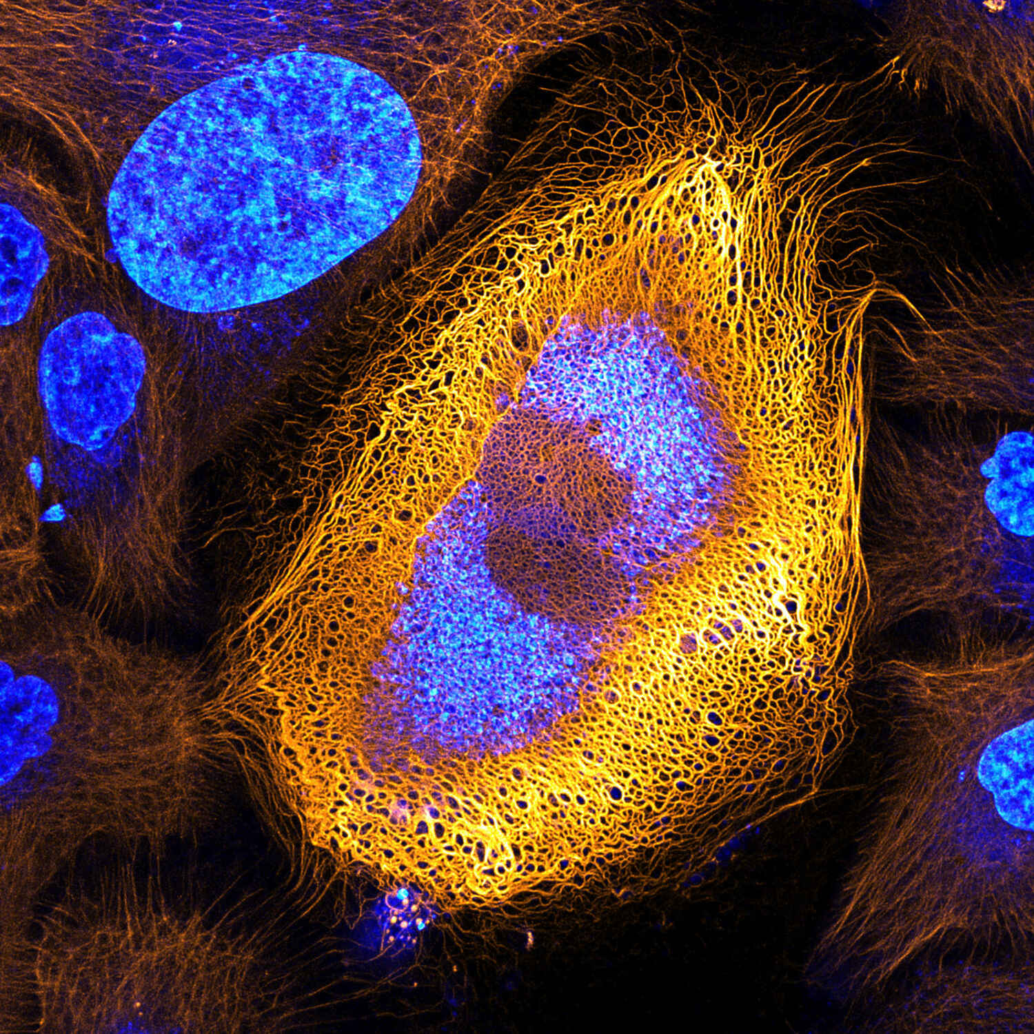
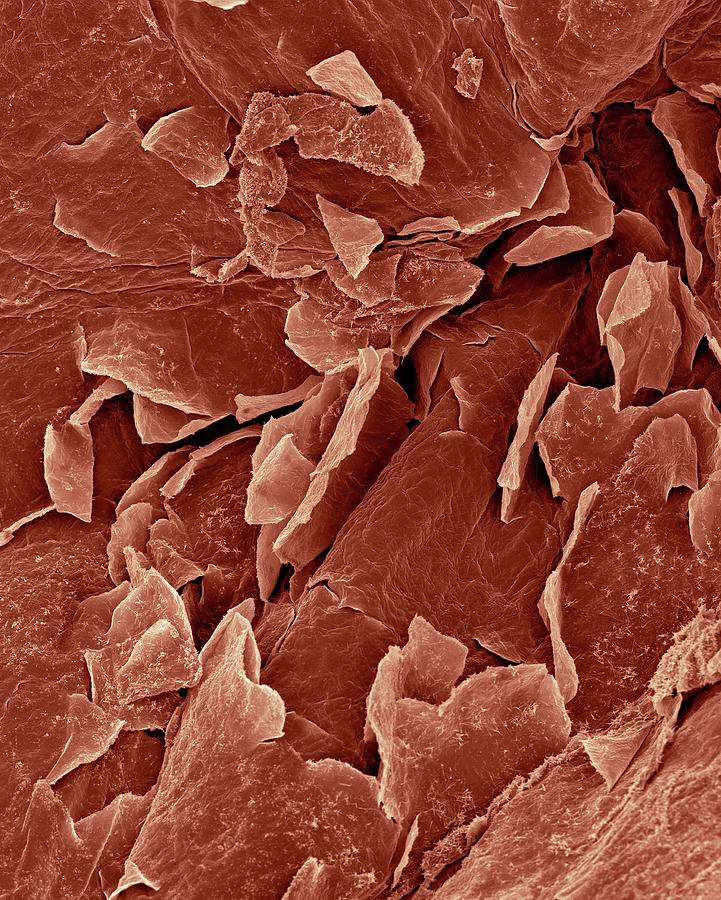
Stunning Microscopic View Of Human Skin Cells Wins 2017 Nikon Small Final thoughts. depending on the strength of the microscope you use, the skin can look like a landscape of layered scales or, with a weaker microscope, a surface that’s crisscrossed with lines and dotted with pores. hairs will appear large and thick, and if you’re lucky, you may even spot a few skin mites! but, as alien as our skin close. Professor susan anderson shows you the microscopic structure of the largest organ in the body the skin. all you need to know about the structure and the ce.

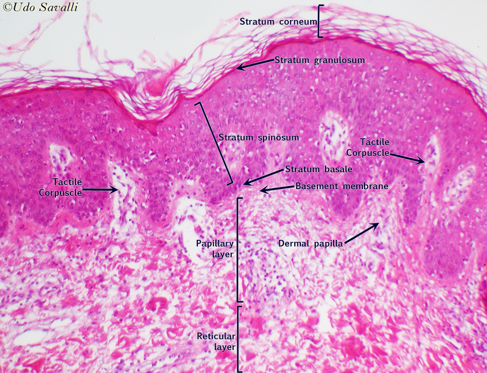
Human Skin Layers Microscope Skin under microscope. skin is the single heaviest organ covering the body surface and directly contacting the external environment. you will find two types of skin – thin or hairy skin and thick or non hairy skin. the skin over the dorsal surface and on the lateral surface of the body is thick. This article will describe the anatomy and histology of the skin. undoubtedly, the skin is the largest organ in the human body; literally covering you from head to toe. the organ constitutes almost 8 20% of body mass and has a surface area of approximately 1.6 to 1.8 m2, in an adult. it is comprised of three major layers: epidermis, dermis and. The skin is the largest organ in the body, covering its entire external surface. the skin has 3 layers—the epidermis, dermis, and hypodermis, which have different anatomical structures and functions (see image. cross section, layers of the skin). the skin's structure comprises an intricate network that serves as the body's initial barrier against pathogens, ultraviolet (uv) light, chemicals. Skin is part of the integumentary system and considered to be the largest organ of the human body. there are three main layers of skin: the epidermis, the dermis, and the hypodermis (subcutaneous fat). the focus of this topic is on the epidermal and dermal layers of skin. skin appendages such as sweat glands, hair follicles, and sebaceous glands are reviewed in depth elsewhere.[1].

Human Skin Epidermis Photograph By Dennis Kunkel Microscopy Science The skin is the largest organ in the body, covering its entire external surface. the skin has 3 layers—the epidermis, dermis, and hypodermis, which have different anatomical structures and functions (see image. cross section, layers of the skin). the skin's structure comprises an intricate network that serves as the body's initial barrier against pathogens, ultraviolet (uv) light, chemicals. Skin is part of the integumentary system and considered to be the largest organ of the human body. there are three main layers of skin: the epidermis, the dermis, and the hypodermis (subcutaneous fat). the focus of this topic is on the epidermal and dermal layers of skin. skin appendages such as sweat glands, hair follicles, and sebaceous glands are reviewed in depth elsewhere.[1]. 1. introduction. the skin is the largest organ of the human body, with an average surface area of 30 m 2 in adults [].as the outer layer of the body—with a thickness of 2–3 mm—it functions as both a physical barrier, protecting its interior against the negative influence of various environmental factors, and an immunological barrier, reducing the effects of injuries and infections. Abstract. this chapter is intended to be a guide to skin studies dealing with the analysis of subcellular structures of cells and tissues by providing an exhaustive sample preparation protocol for transmission electron microscopy (tem). critical steps to obtain good ultrastructure are mentioned as well as notes to improve the sample collection.

Human Skin Outermost Layer Microscopic Photography Science Images 1. introduction. the skin is the largest organ of the human body, with an average surface area of 30 m 2 in adults [].as the outer layer of the body—with a thickness of 2–3 mm—it functions as both a physical barrier, protecting its interior against the negative influence of various environmental factors, and an immunological barrier, reducing the effects of injuries and infections. Abstract. this chapter is intended to be a guide to skin studies dealing with the analysis of subcellular structures of cells and tissues by providing an exhaustive sample preparation protocol for transmission electron microscopy (tem). critical steps to obtain good ultrastructure are mentioned as well as notes to improve the sample collection.

Human Skin Seen Under A Microscope Stock Image C019 9094 Science

Comments are closed.