Parts Of A Neuron And Their Functions With Labelled Diagram

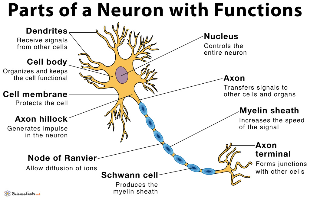
Parts Of A Neuron And Their Functions With Labelled Diagram While they have the common features of a typical cell, they are structurally and functionally unique from other cells in many ways. all neurons have three main parts: 1) dendrites, 2) cell body or soma, and 3) axons. besides the three major parts, there is the presence of axon terminal and synapse at the end of the neuron. As such, neurons typically consist of four main functional parts which include the: receptive part (dendrites), which receive and conduct electrical signals toward the cell body. integrative part (usually equated with the cell body soma), containing the nucleus and most of the cell's organelles, acting as the trophic center of the entire neuron.

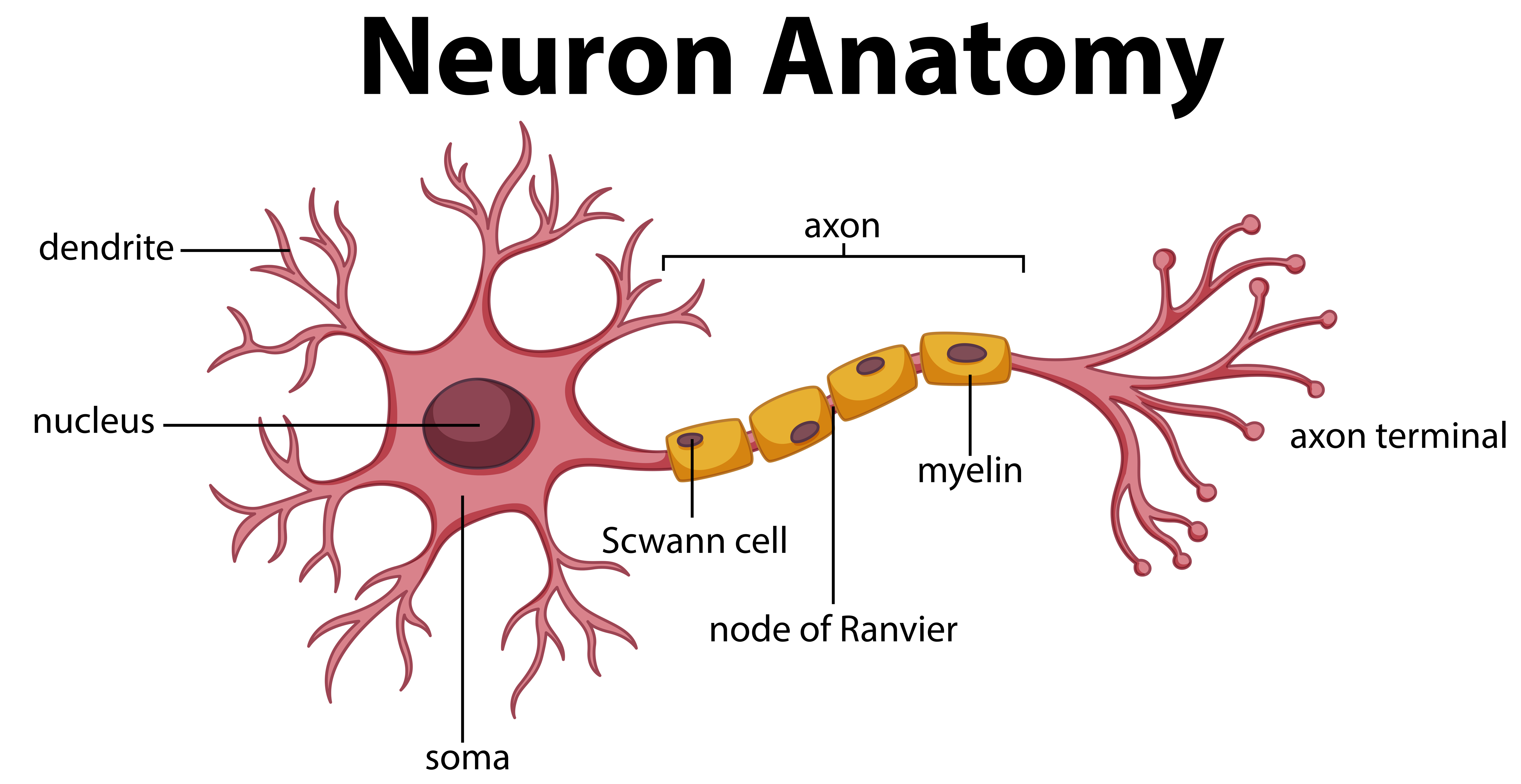
Diagram Of Neuron Anatomy 358962 Vector Art At Vecteezy A neuron is a nerve cell that processes and transmits information through electrical and chemical signals in the nervous system. neurons consist of a cell body, dendrites (which receive signals), and an axon (which sends signals). synaptic connections allow communication between neurons, facilitating the relay of information throughout the body. Here is the description of human neuron along with the diagram of the neuron and their parts. the neuron is a specialized and individual cell, which is also known as the nerve cell. a group of neurons forms a nerve. dendrites–a branch like structure that functions by receiving messages from other neurons and allow the transmission of messages. Neuron structure. figure \(\pageindex{2}\) shows the structure of a typical neuron. the main parts of a neuron are labeled in the figure and described below. figure \(\pageindex{2}\): somatic motor neuron with cell body, axon, axon, myelin sheath, nodes of ranvier, axon terminal, dendrites, synaptic end of the bulbs, and other associated. All labeling quizzes focus on basic anatomy knowledge: recognizing organs and memorizing their names. mastering these essentials is the first step in learning complex anatomy subject matter. structure of the neuron. free quiz neuron labeling quiz for students biology, anatomy and physiology.

Neurons Nerve Cells Structure Function Types Neuron structure. figure \(\pageindex{2}\) shows the structure of a typical neuron. the main parts of a neuron are labeled in the figure and described below. figure \(\pageindex{2}\): somatic motor neuron with cell body, axon, axon, myelin sheath, nodes of ranvier, axon terminal, dendrites, synaptic end of the bulbs, and other associated. All labeling quizzes focus on basic anatomy knowledge: recognizing organs and memorizing their names. mastering these essentials is the first step in learning complex anatomy subject matter. structure of the neuron. free quiz neuron labeling quiz for students biology, anatomy and physiology. Terminal buttons are found at the end of the axon, below the myelin sheath, and are responsible for sending the signal on to other neurons. at the end of the terminal button is a gap known as a synapse. neurotransmitters carry signals across the synapse to other neurons. when an electrical signal reaches the terminal buttons, neurotransmitters. Multipolar neurons have one axon and many dendritic branches. these carry signals from the central nervous system to other parts of your body such as your muscles and glands. unipolar neurons are also known as sensory neurons. they have one axon and one dendrite branching off in opposite directions from the cell body.

Comments are closed.