Three Dimensional Speckle Tracking Imaging Of The Left Ventricleођ

Three Dimensional Speckle Tracking Imaging Of The Left Ventr Three dimensional (3d) speckle tracking echocardiography (3dste) is an advanced imaging technique designed for left ventricular (lv) myocardial deformation analysis based on 3d data sets. 3dste has the potential to overcome some of the intrinsic limitations of two dimensional ste (2dste) in the assessment of complex lv myocardial mechanics. Introduction. three dimensional (3d) speckle tracking echocardiography (ste) combines the advantages of ste and volumetric 3d echocardiography, which shows the left ventricle (lv) in 3d during the cardiac cycle and is also suitable for accurate strain measurements in addition to volumetric assessments using the same virtual 3d lv cast. the present study aimed to confirm the prognostic impact.

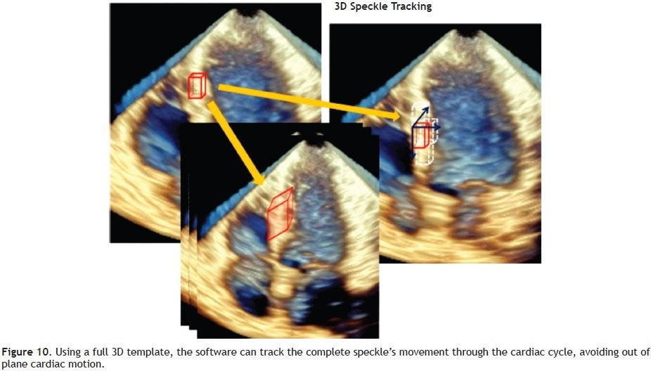
Three Dimensional Speckle Tracking Imaging Of The Left Ventr With the emergence of 2d sti, the abovementioned left ventricular myocardial strain parameters can be measured quantitatively. 3d sti combines real time three dimensional echocardiography and sti and can track myocardial tissue signals in three dimensional space without the limitation of planes, which compensates for the deficiency of 2d sti. Two dimensional strain is a technique of quantifying myocardial deformation from continuous frame by frame tracking of acoustic speckles, which is angle independent (provides measurements of three deformation components: longitudinal, radial and circumferential), less subject to artefacts, and simpler to obtain than doppler derived tissue. Three dimensional speckle tracking imaging (3d sti) is a cutting edge approach that utilizes 3d echocardiography and speckle technology to track myocardial tissue speckles [citation 11]. by employing 3d sti, it becomes possible to assess left ventricular systolic function through the measurement of parameters such as radial, longitudinal, and. Larly, images of speckle tracking on the 2 dimensional b mode images are modeled after those in (a). (c) the 3 di mensional speckle tracking method of the volume of interest (red line cubic template) from one volume (starting vol ume) to the next volume (white dashed line cubic template).

Function And Mechanics Of The Left Ventricle From Tissue Doppler Three dimensional speckle tracking imaging (3d sti) is a cutting edge approach that utilizes 3d echocardiography and speckle technology to track myocardial tissue speckles [citation 11]. by employing 3d sti, it becomes possible to assess left ventricular systolic function through the measurement of parameters such as radial, longitudinal, and. Larly, images of speckle tracking on the 2 dimensional b mode images are modeled after those in (a). (c) the 3 di mensional speckle tracking method of the volume of interest (red line cubic template) from one volume (starting vol ume) to the next volume (white dashed line cubic template). With the arrival of three dimensional systems, the entire left ventricle can be evaluated with this technique, lacking the inherent weakness of two dimensional and tissue doppler methods. three dimensional speckle tracking (3dst) has potential to be an ideal tool to assess not only global myocardial function but regional function through. To investigate the relationship between left ventricular (lv) myocardial mechanics evaluated by three dimensional speckle tracking echocardiography (3d ste) and degree of coronary artery stenosis in patients with coronary artery disease (cad). ninety seven suspected cad patients without lv regional wall motion abnormality (rwma) observed visually form traditional echocardiography were divided.

Function And Mechanics Of The Left Ventricle From Tissue Doppler With the arrival of three dimensional systems, the entire left ventricle can be evaluated with this technique, lacking the inherent weakness of two dimensional and tissue doppler methods. three dimensional speckle tracking (3dst) has potential to be an ideal tool to assess not only global myocardial function but regional function through. To investigate the relationship between left ventricular (lv) myocardial mechanics evaluated by three dimensional speckle tracking echocardiography (3d ste) and degree of coronary artery stenosis in patients with coronary artery disease (cad). ninety seven suspected cad patients without lv regional wall motion abnormality (rwma) observed visually form traditional echocardiography were divided.

Analysis Report Of Left Ventricle By Three Dimensional Speckle Trac

Comments are closed.