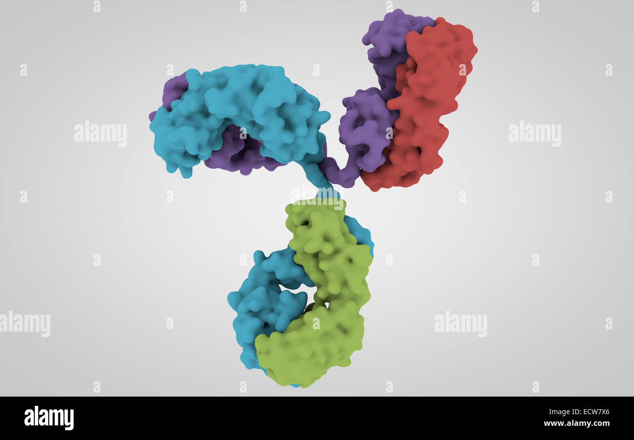
Threedimensional Structure Antibody Molecule Stock Illustration 806873

Threedimensional Structure Antibody Molecule Stock Illustration 806873 Find threedimensional structure antibody molecule stock images in hd and millions of other royalty free stock photos, illustrations and vectors in the shutterstock collection. thousands of new, high quality pictures added every day. The 2.8 a resolution three dimensional structure of a complex between an antigen (lysozyme) and the fab fragment from a monoclonal antibody against lysozyme has been determined and refined by x ray crystallographic techniques. no conformational changes can be observed in the tertiary structure of lysozyme compared with that determined in native.

3d Structure Of Antibody Stock Photo Alamy Figure 4. a 3d image of an individual particle of igg1 antibody reconstructed by individual particle electron tomography (ipet). (a) schematics of the et imaging of an individual particle of protein from a series of tilt angles. (b) the step by step process of the ipet 3d structure of an individual particle of igg1. 1.1. overall features of the immunoglobulin. the intact antibody molecule shown in figure 1 has three functional components, two fragment antigen binding domains (fabs) and the fragment crystallizable (fc), with the two fabs linked to the fc by a hinge region that allows the fabs a large degree of conformation flexibility relative to the fc. The structure of the complex between hen egg white lysozyme and the fab of a monoclonal anti lysozyme antibody (d1.3) shows that the combining site of antibodies is not merely a cleft delineated. Nature immunology 17, s11 (2016) cite this article. the characteristic y shape of antibodies is arguably the most easily recognizable protein structure. since the 1970s, x ray crystallography and.

Structure Of An Antibody Stock Illustration Illustration Of The structure of the complex between hen egg white lysozyme and the fab of a monoclonal anti lysozyme antibody (d1.3) shows that the combining site of antibodies is not merely a cleft delineated. Nature immunology 17, s11 (2016) cite this article. the characteristic y shape of antibodies is arguably the most easily recognizable protein structure. since the 1970s, x ray crystallography and. The structure of a typical antibody molecule. antibodies are the secreted form of the b cell receptor. an antibody is identical to the b cell receptor of the cell that secretes it except for a small portion of the c terminus of the heavy chain constant region. in the case of the b cell receptor the c terminus is a hydrophobic membrane anchoring. Disulfide bond illustration on the three dimensional structure of an antibody (pdb id: 1ad9). the light chain (chain a) was marked in white while the heavy chain (chain b) was marked in green.

The Structure Of A Typical Antibody Molecule Antibodies And Amino Acids The structure of a typical antibody molecule. antibodies are the secreted form of the b cell receptor. an antibody is identical to the b cell receptor of the cell that secretes it except for a small portion of the c terminus of the heavy chain constant region. in the case of the b cell receptor the c terminus is a hydrophobic membrane anchoring. Disulfide bond illustration on the three dimensional structure of an antibody (pdb id: 1ad9). the light chain (chain a) was marked in white while the heavy chain (chain b) was marked in green.

Comments are closed.