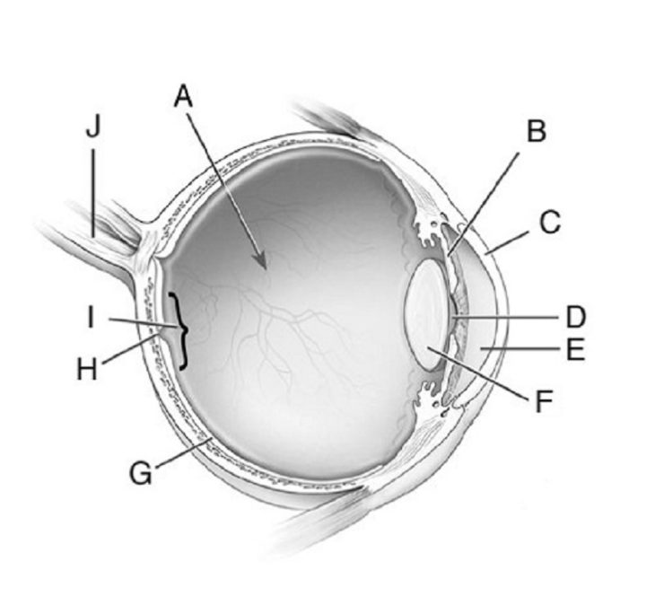
Unlabeled Diagram Of The Eye

Printable Eye Diagram Quiz Unlabeled 101 Diagrams Labelling the eye. use this interactive to label different parts of the human eye. drag and drop the text labels onto the boxes next to the diagram. selecting or hovering over a box will highlight each area in the diagram. the human eye has several structures that enable entering light energy to be converted to electrochemical energy. Behind the anterior chamber is the eye’s iris (the colored part of the eye) and the dark hole in the middle called the pupil. muscles in the iris dilate (widen) or constrict (narrow) the pupil to control the amount of light reaching the back of the eye. directly behind the pupil sits the lens. the lens focuses light toward the back of the eye.

Diagram Of An Eye Unlabeled Human Eye Diagram Human Eye Eyesо Diagram of visual projection pathway from eyes to brain (visual cortex), unlabeled. astigmatism, labeled diagram. the oval shape of the cornea of the eye in people with astigmatism creates multiple focal points. Take a look at the diagram of the eyeball above. here you can see all of the main structures in this area. spend some time reviewing the name and location of each one, then try to label the eye yourself without peeking! using the eye diagram (blank) below. unlabeled diagram of the eye. click below to download our free unlabeled diagram of. Eye diagrams. the bony orbit. field defects. anterior chamber angle and ciliary body. fundus diagram. quiz on the 5 layers of the cornea. the retina at its junction with the optic disc. brainstem anatomy of the cranial nerves involved in eye movement. Iris: the iris is the colored part of the eye that regulates the amount of light entering the eye. lens: the lens is a clear part of the eye behind the iris that helps to focus light, or an image, on the retina. macula: the macula is the small, sensitive area of the retina that gives central vision. it is located in the center of the retina.

Comments are closed.